
Fig. 2.
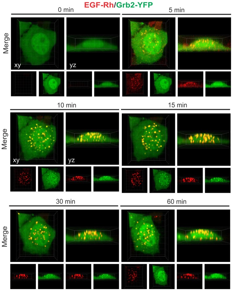
Time-lapse imaging of Grb2–YFP in cells stimulated with 2 ng/ml EGF–Rh (0–60 minutes). 3D imaging of HeLa-Grb2–YFP cells incubated with 2 ng/ml EGF–Rh at 37°C for the indicated times, as described in the Materials and Methods. Selected x–y and x–z images are presented. See corresponding supplementary material Movies 2 and 3.