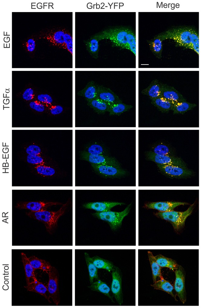
Fig. 6.

Colocalization of Grb2–YFP with EGFR in HeLa-Grb2–YFP cells treated with unlabeled EGFR ligands. Cells grown on glass coverslips were left untreated (Control) or treated with EGF (2 ng/ml), TGFα (2 ng/ml), HB-EGF (2 ng/ml) or AR (400 ng/ml) for 30 minutes at 37°C, fixed and stained with mouse monoclonal EGFR (mAb528) followed by secondary antibody conjugated with Cy3. Cell nuclei were stained with DAPI. Scale bar: 10 µm.
