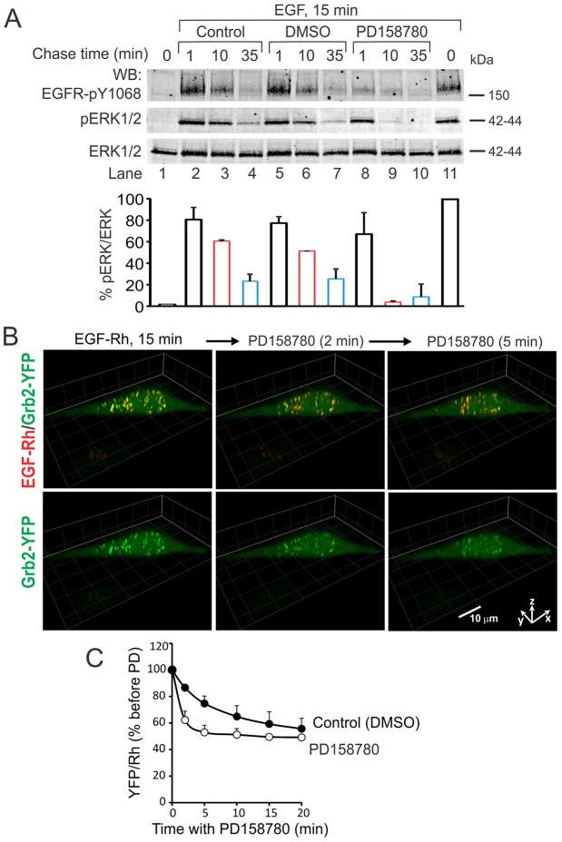
Fig. 8.
Effect of the EGFR kinase inhibitor on Grb2–YFP localization in endosomes and ERK1/2 activity. (A) HeLa-Grb2–YFP cells were incubated (lanes 2–11) or not (lane 1) with EGF (40 ng/ml) for 15 minutes at 37°C. The incubation (Chase) was continued in the same medium (Control, lanes 2–4), or after addition of DMSO (vehicle, lanes 5–7) or PD158780 (50 nM final concentration, lanes 8–10) for additional 1, 10 and 35 minutes. Cell lysates were probed by western blotting with antibodies to pTyr1068 of EGFR (pEGFR), phospho-ERK1/2 and ERK1/2. Bars below blots represent quantification of the ERK1/2 phosphorylation normalized to total ERK1/2 (pERK/ERK), and expressed as a percent of the pERK:ERK ratio in cells treated with EGF for 15 minutes (lane 11). The data are averaged from two independent experiments. (B) HeLa-Grb2–YFP cells were incubated with EGF–Rh (4 ng/ml) for 15 minutes at 37°C. DMSO (vehicle) or PD158780 were added to EGF–Rh-containing medium, and the cells were further incubated for 0–20 minutes at 37°C. 3D time-lapse imaging (26 planes, 500 nm intervals) was performed before and after injecting PD158780. Selected time-lapse 3D images are presented. Fluorescence intensity ranges of all images are identical. Scale bar: 10 µm. (C) Quantification of the ratio of YFP and Rhodamine fluorescence intensities in endosomes of cells treated with DMSO or PD158780 as in B was performed as in Fig. 4A. Values are means (± s.e.m.) of YFP:Rhodamine ratios from four to eight cells with similar expression levels of Grb2–YFP.

