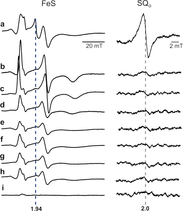
Figure 2.

Testing the sensitivity of g = 1.94 and g = 2.0 signals to inhibitors and mutations that abolish the activity of the Qo site. X-Band EPR spectra of isolated WT cytochrome bc1 obtained under the conditions described for Figure 1c in the presence of antimycin alone (a) or antimycin and one of the Qo site inhibitors: tridecyl-stigmatellin (b), atovaquone (c), famoxadone (d), myxothiazol (e), azoxystrobin (f), or kresoxim-methyl (g). Spectra of antimycin-inhibited mutants G158W (h) and the b-c1 subcomplex (i). The left panel (FeS) shows spectra measured at 20 K in a magnetic field range of the FeS signal, and the right panel (SQo) shows spectra measured at 200 K in a magnetic field range typical of organic radicals.
