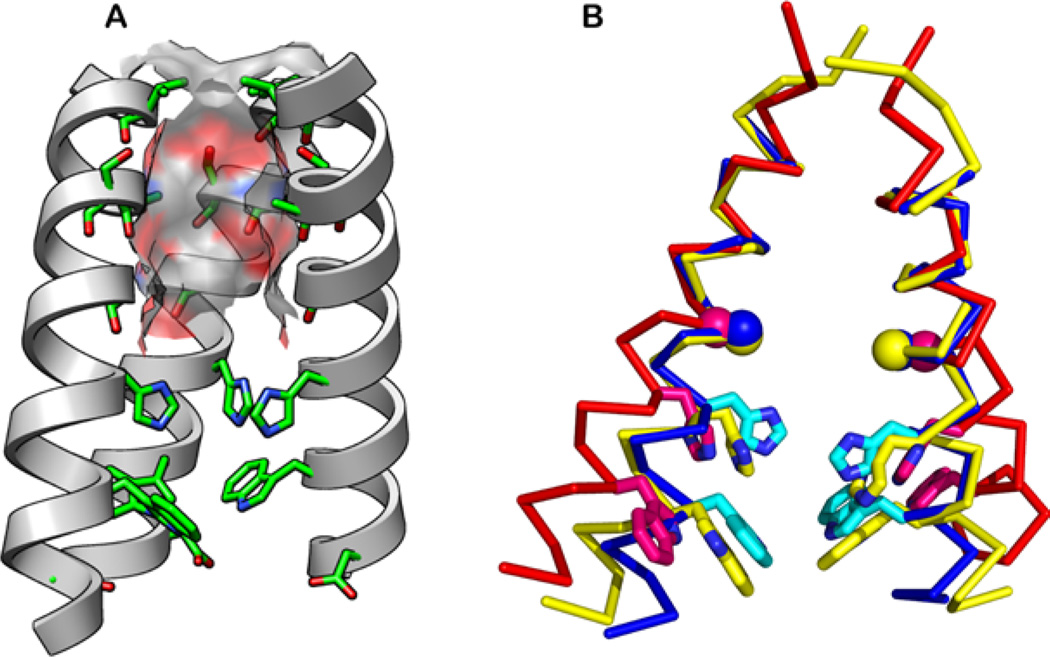
Figure 2.
Structures of the TM domain of M2. (A) The N-terminal pore is lined by the hydroxyl of Ser31 and backbone carbonyl groups (pictured structure is the 1.65Å crystal structure at pH 6.5, PDB: 3LBW). The molecular surface of the channel is color-coded with the oxygen atoms in red, carbon in gray, and nitrogen in blue. The “front” helix has been removed for clarity. (B) Superimposed solid-state NMR structure at pH 7.5 (2L0J, yellow), the 1.65-Å crystal structure at pH 6.5 (3LBW, blue), and the 3.5-Å crystal structure at pH 5.3 (3C9J, red). His37 and Trp41 side chains (sticks) and Gly34 Cα (ball) are shown.

