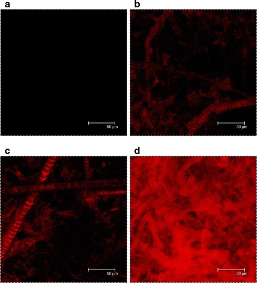
Fig. 5.

Confocal micrographs of nude mouse skin at an original magnification of ×630 with non-treatment (blank) (a), in vivo topical administration of aqueous control (b), squarticles in NLC type (c), and squarticles in NE type (d) containing Nile red (0.1%) as a dye for 6 h. The image is a summary of 15 fragments at various skin depths
