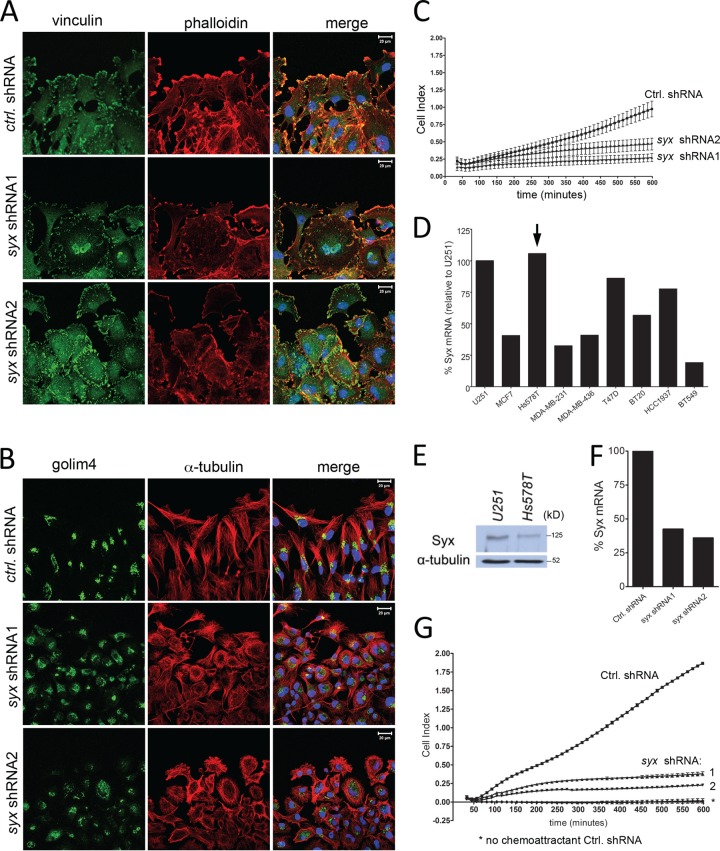
Fig 2.
Syx is required for tumor cell polarization and directional cell migration. (A) Immunofluorescence staining of vinculin and f-actin (phalloidin) at the leading edge of a wounded U251 monolayer. Cells were fixed and processed for staining 5 h after wounding. (B) As in panel A, cells were fixed and stained for Golgi integral membrane protein 4 (golim4) and α-tubulin 5 h after wounding was induced. Note the complete loss of directional Golgi staining in Syx-depleted cells. (C) xCELLigence Transwell migration assay measuring the real-time migration pattern of control shRNA versus syx shRNA expressing U251 cells toward the FBS chemoattractant. (D) qRT-PCR analysis of Syx mRNA levels in breast cancer cell lines, normalized to respective 18S rRNA levels. The results were then calculated as a percentage of 18S-normalized Syx mRNA in U251 cells. (E) Western blot analysis showing the expression level of Syx protein in Hs578T cells compared to U251. (F) qRT-PCR analysis of Syx mRNA levels in Hs578T cells expressing control shRNA versus either of two nonoverlapping syx shRNAs, normalized to GAPDH mRNA levels. (G) As in panel C, the directional migration of syx shRNA expressing Hs578T cells toward a gradient of FBS was impaired.

