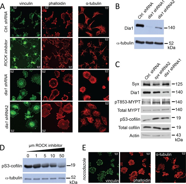
Fig 4.
Dia1 opposes ROCK and phenocopies Syx in U251 cell morphology. (A) Immunofluorescence experiment comparing the morphology of U251 cells expressing control versus either of two nonoverlapping dia1 shRNAs, or treated with ROCK inhibitor (Y-27632, 10 μM, 12 h). Fixed cells were stained for vinculin and f-actin (phalloidin) or for α-tubulin. ROCK inhibition resulted in a stellate, multipolar cell morphology, whereas dia1 shRNA expression induced a well-spread, apolar phenotype. (B) Western blot analysis showing the efficiency of Dia1 protein depletion in U251 cells expressing the dia1 shRNAs. (C) Western blot analysis of phosphoT853-MYPT, total MYPT, phosphoS3-cofilin, and total cofilin in U251 cells expressing control-, syx-, or dia1-specific shRNAs. (D) Western blot analysis of phosphoS3-cofilin in U251 cells treated overnight with increasing amounts of ROCK inhibitor (Y-27632). (E) U251 cells were treated with nocodazole (10 μM, 5 h), fixed, and stained for vinculin and f-actin (phalloidin) or for α-tubulin. Nocodazole treatment increased focal adhesions and induced an apolar cell morphology.

