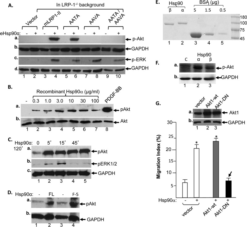
Fig 6.
The NPVY motif connects eHsp90α signaling to the Akt but not ERK1/2 pathway. (A) Based on the results of the protein kinase array, the endogenous LRP-1-downregulated HDFs expressing mLRP1-II with the AATA, AAVA, or AATA/AAVA mutations were serum starved and stimulated without or with eHsp90α (0.1 μM, 15 min.). Protein-equalized lysates of the cells were subjected to Western blotting with anti-phospho-Akt(S437), anti-phospho-ERK1/2, or control antibody. (B) Parental HDFs were serum starved and treated without or with increasing concentrations of eHsp90α (15 min) or PDGF-BB (50 ng/ml, 5 min). Total lysates were subjected to Western blotting with anti-phospho-Akt(S437) or anti-Akt protein antibody. (C) Parental HDFs were serum starved and treated without or with eHsp90α (10 μg/ml or 0.1 μM) for the indicated time. Total lysates were subjected to Western blotting with anti-phospho-Akt(S437), anti-phospho-ERK1/2, or anti-GAPDH antibody. (D) Parental HDFs were serum starved and stimulated with either full-length eHsp90α (0.1 μM) or F-5 (0.1 μM) for 15 min. Total lysates were immunoblotted with anti-phospho-Akt(S437) (a) or anti-GAPDH antibody (b). (E) Demonstration of the FPLC-purified human recombinant Hsp90β (lane 1) and Hsp90α (lane 2) proteins. (F) Parental HDFs were serum starved and stimulated with equal amounts of the Hsp90α or Hsp90β protein, and activation of Akt was measured by using anti-phospho-Akt antibody (a) or GAPDH antibody (b). (G) HDFs were infected with lentiviral vector carrying either the wt (lane 2) or dominant-negative (DN) mutant Akt1 gene. The inset shows a Western blot of the expressed Akt gene products. The bar graph shows that the cells then were subjected to a colloidal gold migration assay (see Materials and Methods). Only the migration index is shown.

