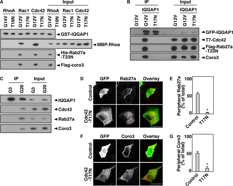
Fig 4.
Cdc42 regulates the interaction between IQGAP1 and GDP-bound Rab27a. (A) An in vitro binding assay was performed using purified bead-bound GST-IQGAP1, His-Rab27a-T23N, Flag-coronin 3, and an MBP-tagged RhoA, Rac1, or Cdc42 mutant. Following incubation, bead-bound proteins were analyzed by immunoblotting using anti-GST, anti-MBP, anti-His, and anti-Flag antibodies. (B) COS-7 extracts expressing GFP-IQGAP1, Flag-Rab27a-T23N, and one of the T7-Cdc42 mutants (the G12V or T17N mutant) were immunoprecipitated with an anti-GFP antibody. The immunocomplexes were analyzed by immunoblotting using anti-GFP, anti-T7, anti-Flag, and anti-coronin 3 antibodies. The percentage of input protein coimmunoprecipitated was 0.2%. (C) MIN6 cells were incubated with 3 or 20 mM glucose for 5 min. The cell extracts were immunoprecipitated with an anti-IQGAP1 antibody. The immunocomplexes were analyzed by immunoblotting with anti-IQGAP1, anti-Cdc42, anti-Rab27a, and anti-coronin 3 antibodies. The percentage of input protein coimmunoprecipitated was 0.1%. (D) MIN6 cells expressing Flag-Rab27a together with GFP or GFP-Cdc42-T17N were incubated with 20 mM glucose for 5 min. The cells were immunostained with an anti-Flag (red) antibody. (E) The percentage of cells with a peripheral distribution pattern of Flag-Rab27a was analyzed. (F) MIN6 cells expressing Flag-coronin 3 together with GFP or GFP-Cdc42-T17N were incubated with 20 mM glucose for 5 min. The cells were immunostained with an anti-Flag (red) antibody. Bars, 10 μm. (G) The percentage of cells with a peripheral distribution pattern of Flag-coronin 3 was analyzed. Peripheral distribution and statistical analyses were performed as described in the legend to Fig. 3C.

