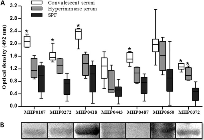
Fig 2.

Antigenicity analysis of the recombinant proteins. (A) ELISA of the seven recombinant M. hyopneumoniae proteins against 30 convalescent-phase pig serum samples, four hyperimmune pig serum samples (experimentally infected with M. hyopneumoniae 7448), and 10 SPF pig sera (all sera were diluted 1:100). Peroxidase-conjugated anti-pig IgG (diluted 1:2,000) was used as the secondary antibody. Asterisks indicate significant differences compared to SPF sera using the Tukey test with GraphPad Prism 4 software (P < 0.01). Boxes represent the interquartile range in the middle 50% of the absorbance values. Whiskers represent the minimum and maximum values. (B) Western blot analysis of the recombinant proteins probed with the pooled sera of 5 convalescing pigs (diluted 1:100). Peroxidase-conjugated anti-pig IgG (diluted 1:2,000) was used as the secondary antibody.
