Fig 3.
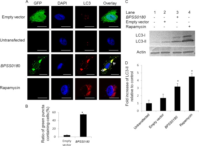
BPSS0180 induces autophagy in HeLa cells. (A) Representative confocal images of HeLa cells transfected with empty vector (green) or GFP-tagged BPSS0180 (green), untransfected cells (untreated), and cells treated with rapamycin. Cells were fixed, permeabilized, and stained for nuclei (blue) or LC3 (red). Arrows indicate colocalization of punctate structures with LC3, defined by the presence of green punctate structures in BPSS0180-transfected cells (green) which were fully overlaid by intense red. Bars, 10 μm. (B) Quantitative analysis of green puncta induced by empty vector or BPSS0180 in HeLa cells. (C) Western blots. (Top) LC3 expression of HeLa cells transfected with empty vector or BPSS0180, untransfected cells (untreated), or cells treated with rapamycin. (Bottom) Actin as a loading control. (D) Fold increases in LC3-II were determined by densitometric quantitative comparison of each LC3-II band to the same band in untransfected control cells (lane 2). Each band was normalized against the actin control band. Asterisks indicate P values of <0.05 relative to untransfected control cells.

