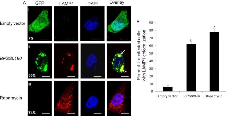
Fig 4.
BPSS0180-induced puncta colocalize with the lysosome marker LAMP1. (A) Representative confocal images of HeLa cells transfected with empty vector (green) or GFP-tagged BPSS0180 (green) or left untransfected and treated with rapamycin. Cells were fixed, permeabilized, and stained for nuclei (blue) and LAMP1 (red), a marker of lysosomes. Arrows indicate cells with punctate structures colocalizing with LAMP1. Colocalization of puncta with LAMP1 was defined by the proportion of green puncta overlapping regions of anti-LAMP1 staining. Bars, 10 μm. (B) Quantitative analysis of cells with LAMP1 punctate structures. Asterisks indicate P values of <0.05 relative to cells transfected with empty vector.

