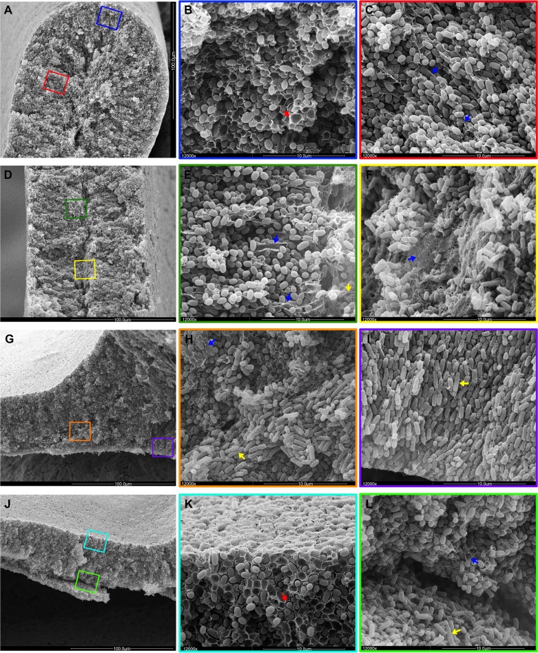
Fig 7.
Spatial arrangement of cellulose, curli and flagella and cell physiology in the context of the two-layer biofilm architecture. For an overview, low-magnification SEM images of AR3110 cross-sections at the tip (A), middle body (D), and base (G) of a ridge and a flat area (J) are shown in the left column of images. The middle and right columns show color-boxed images at ×12,000 magnification corresponding to the color-boxed zones in the overview images. A red arrow in panel B points to basket-like matrix structure typical for curli fiber networks. Blue arrows in panels C, E, F, and H label sheet-like material indicative of cellulose. A yellow arrow in panel E points to flagellum-like filaments in the inner region of the ridge characterized by elongated growing cells. Yellow arrows in panels H and I point to rod-shaped bacteria in the growth zone below the ridge. A red arrow in panel K points matrix-encased bacteria in the upper layer with starving cells. Blue and yellow arrows in panel L indicate cells in contact with sheet-like and filamentous matrix material at the base of the upper colony layer and elongated rod-shaped cells entangled by flagella in the lower layer, respectively.

