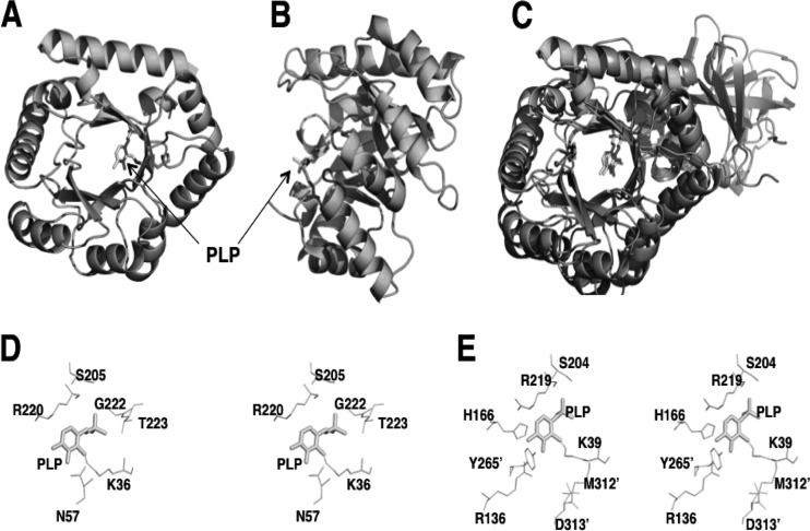
Fig 1.
Structure of YggS and comparison to bacterial AR. A front view (A) and side view (B) of the YggS structure are represented. The PDB code used for YggS is 1W8G. (C) The superposition of the YggS structure onto that of the monomeric structure of G. stearothermophilus AR (PDB entry 1SFT). (D and E) Stereo view of the putative active site of YggS (D) and the active site of AR (E).

