Fig 6.

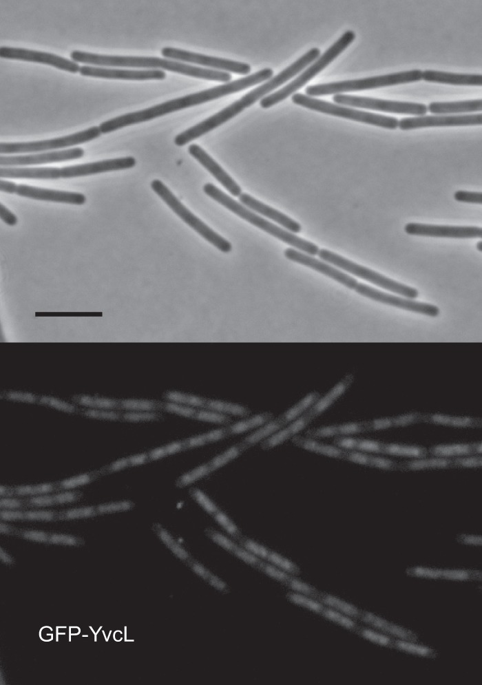
Localization of mGFP-YvcL in B. subtilis cells. Fluorescence microscopy images of strain PG736 containing a Pxyl-mgfp-yvcL fusion and yvcL deletion, grown in LB medium at 30°C in the presence of 0.01% xylose, are shown. Scale bar, 5 μm.

Localization of mGFP-YvcL in B. subtilis cells. Fluorescence microscopy images of strain PG736 containing a Pxyl-mgfp-yvcL fusion and yvcL deletion, grown in LB medium at 30°C in the presence of 0.01% xylose, are shown. Scale bar, 5 μm.