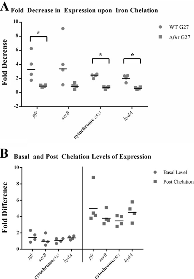
Fig 1.

apo-Fur regulation of the cytochrome c553 gene, hydA, and serB. RNA was isolated from exponential-phase cultures of WT and Δfur strains of H. pylori under iron-replete and iron chelation conditions as detailed in Materials and Methods. Riboprobes for the cytochrome c553 gene, hydA, serB, and the control promoter, pfr, were used to evaluate changes in expression of these genes. The fold decrease in expression upon iron limitation is shown in panel A. The fold difference in the basal levels of gene expression between the Δfur and WT strains is shown on the left side of panel B. The fold difference in the postchelation levels of gene expression between the Δfur and WT strains is shown on the right side of panel B. The geometric means of the fold decrease and fold difference in the relative levels of expression from the four biological replicates are shown as black lines. An asterisk above a bracket indicates that there was a statistically significant difference in the fold decrease between WT and Δfur strains upon iron chelation (P ≤ 0.01).
