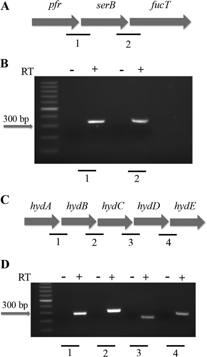
Fig 3.

Organization of the pfr and hydA operons. The genes encoding pfr, serB, and fucT are organized into an operon as shown in panel A. Panel B shows the PCR amplification of the gene junctions between pfr and serB and serB and fucT using cDNA (+) and no-RT (−) control reactions as templates. The organization of the hydACBDE operon is shown in panel C. Panel D shows the PCR amplification of the hydAB, hydBC, hydCD, and hydDE gene junctions using cDNA (+) and no-RT (−) control reactions as templates. The individual junctions are indicated by numbers (A and C), which correspond to the numbers shown below the gel images (B and D). Each junctional PCR was conducted twice using biologically independent cDNA templates, with similar results.
