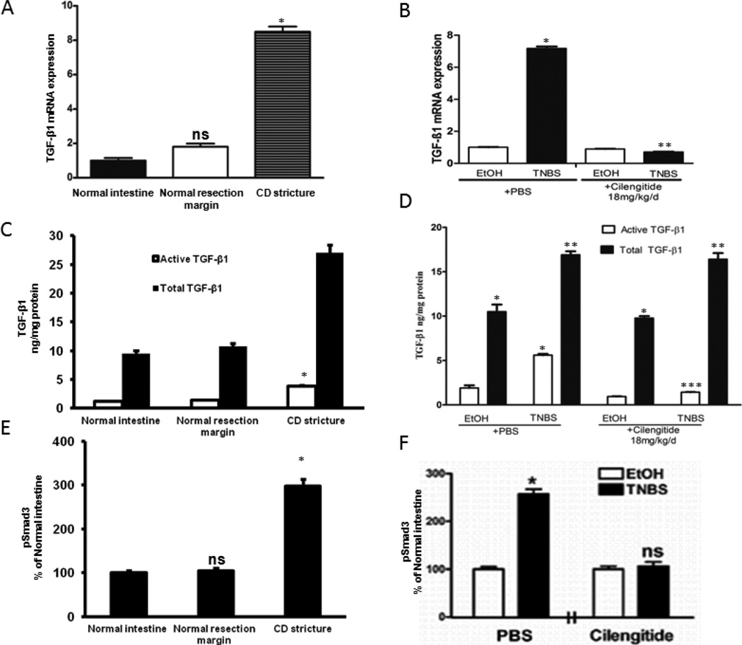
Figure 2. Increased TGF-β1 expression and Smad3 phosphorylation in stricturing Crohn’s disease and in chronic TNBS colitis.
A&B: Increased TGF-β1 transcripts in smooth muscle cells of human strictured intestine compared to normal margin (A) and rat colon after 42 d of chronic TNBS colitis compared to EtOH controls (B). C-F: Tissue levels of active TGF-β1 and of phosphorylated Smad3 in strictured regions of Crohn’s disease in humans (C&E) and after 42 d of chronic TNBS-induced colitis in rats (D&F). Both active TGF-β1 and pSmad3(Ser423/425) levels are αVβ3 RGD-dependent and inhibited by cilengitide. Tissue active and total TGF-β1 levels were measured by ELISA in 9 paired human samples, and in 12 EtOH and TNBS naïve rats or rats treated with PBS or Cilengitide (18 mg/kg/d i.p). * denotes P < 0.05 vs normal margin in humans or EtOH control in rats; ** denotes P < 0.05 vs total TGF-β1 in normal margin in humans, or EtOH/PBS treated rats; *** denotes P < 0.05 vs TNBS/PBS treated rats; ns denotes no significant difference vs TNBS/PBS control rats.

