
Figure 1.
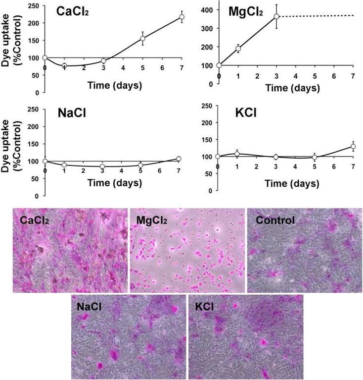
Rose bengal staining in stratified human corneal epithelial cells incubated with 50 mM of individual electrolytes for different time periods. Dashed line in the magnesium chloride graph indicates uniform cell death with longer periods of incubation. Phase contrast images shown below correspond to representative areas following 7-day incubations with or without (control) hypertonic electrolytes.