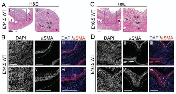
Figure 1. Development of the pylorus between E14.5 and E16.5 involves differentiation of the dorsal OLM and changes in ICM shape.
H&E staining of WT pylorus at (A) E14.5 or (C) E16.5; the dorsal pylorus, within the boxed region in (i) is enlarged in (ii). Immunofluorescence of the dorsal WT pylorus at (B) E14.5 or (D) E16.5: (i,iv) DAPI; (ii,v) αSMA; or (iii,vi) merged. (A–B) Though the ICM is well-differentiated and strongly αSMA positive at E14.5, cells of the OLM stain weakly for αSMA (asterisk in Bvi). (C–D) Expansion and differentiation of the OLM (asterisk in Dvi) is associated with an increase in αSMA expression and inward displacement of the ICM, resulting in pyloric sphincter constriction. For all images, stomach (S) is left; duodenum (D) is right; and, dorsal is top. Green lines mark the epithelial basement membrane, and white lines separate ICM and OLM. E = epithelium; SM = sub-epithelial mesenchyme; and, PM = pancreatic mesenchyme. Scale bars represent 100 μm.

