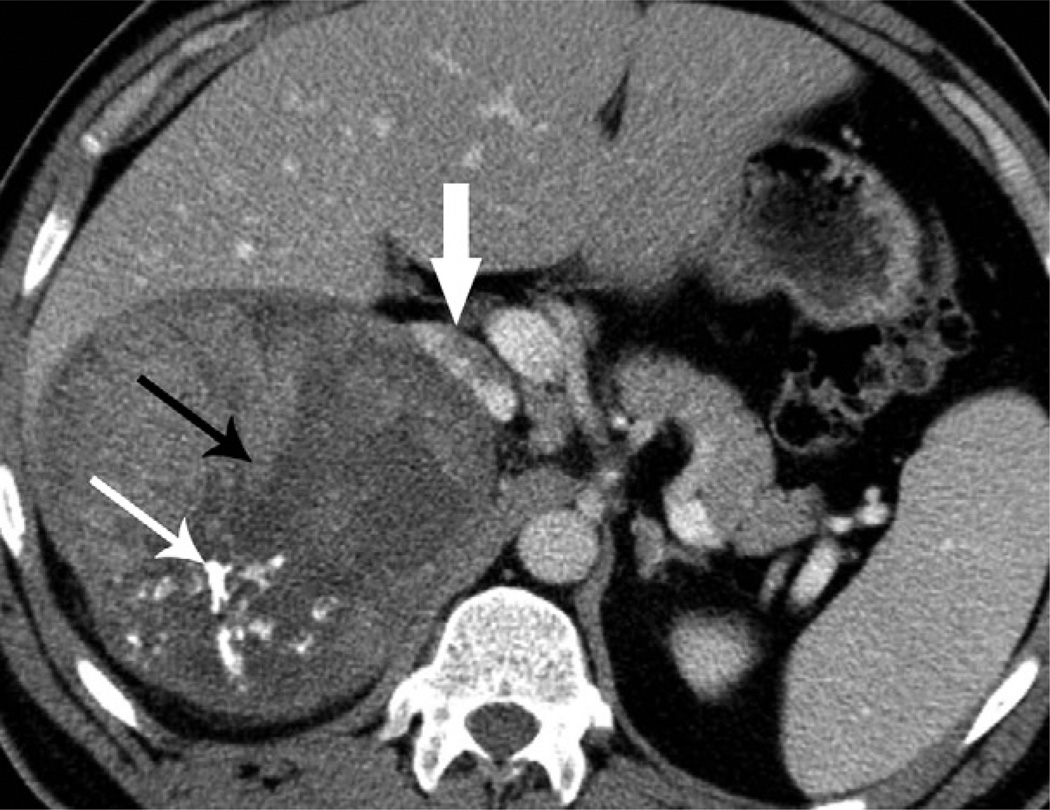
Figure 2.
A 42-year-old man with right ACC. Contrast-enhanced CT image shows a 13 cm, well-circumscribed, lobulate mass with irregular calcifications (thin white arrow) and a central stellate area of low density (thin black arrow). The adjacent liver parenchyma and the IVC (thick white arrow) are displaced anteriorly.

