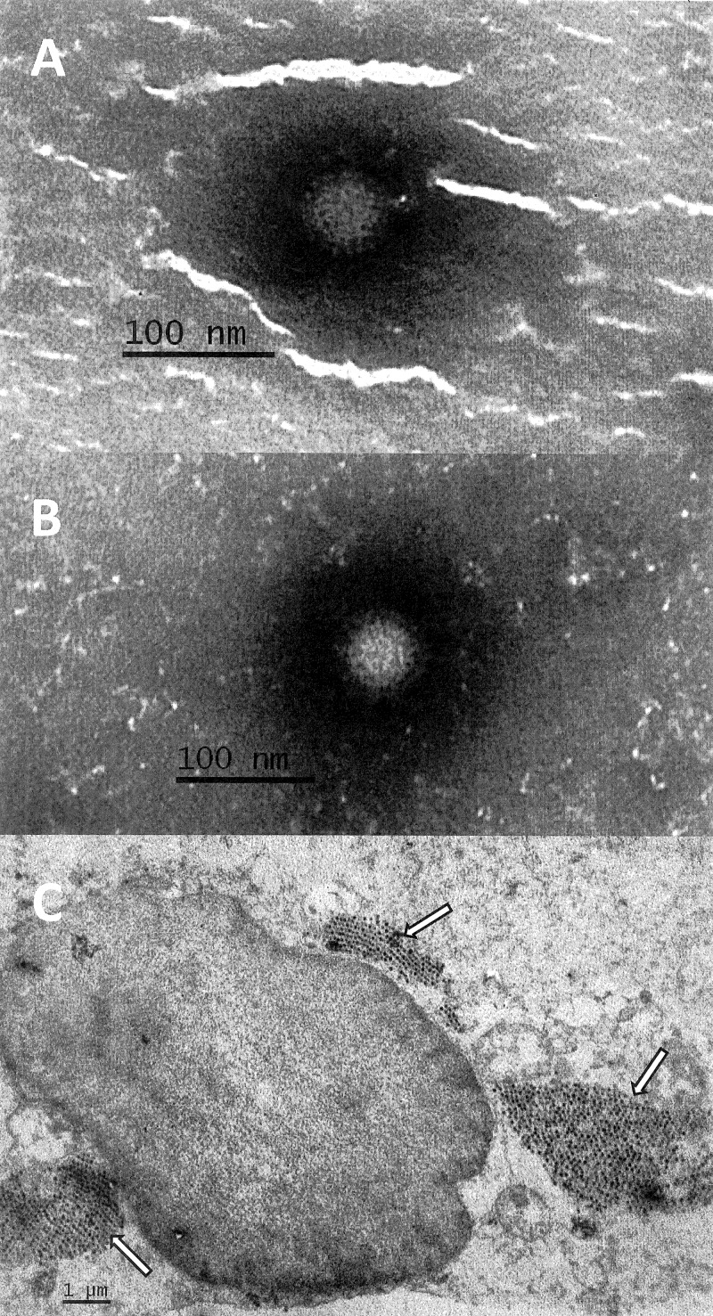
Fig 1.
Electron micrographs of reoviruses in stool suspension (A) (magnification, 100,000×) cell culture supernatant (B) (magnification, 100,000×), and ultrathin section of LLC-MK2 cells (C) (magnification, 10,000×) infected with the SI-MRV01 orthoreovirus strain. Arrows in Fig. 1C indicate reovirus particles.

