
Fig. 1.
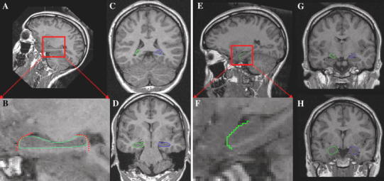
Manual segmentation of the hippocampus and the amygdala. a For the tracing of the hippocampus, each brain was realigned along its longitudinal axis. b Magnification of the sagittal view showing the outlined hippocampus. This step enhances segmentation accuracy in critical coronal slices. c Final coronal segmentation started always at the posterior end of the hippocampus. d Exemplary coronal slice of the hippocampal head. e For the amygdala tracing, the brains remained oriented along the anterior–posterior commissure (AC–PC) line as originally measured. f Magnification of the sagittal view showing how the hippocampal head was separated from the amygdala. g Again, segmentation was conducted from the posterior part to the anterior end of the amygdala. h Exemplary slice of the left and right amygdala. Details on the segmentation protocols can be found in the “Materials and methods” section