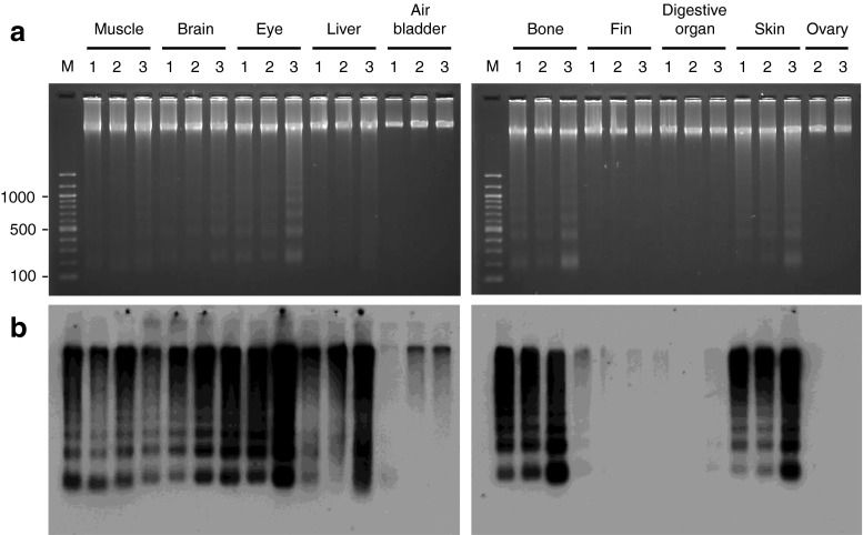
Fig. 7.
Interindividual variations in DNA fragmentation in zebrafish tissues. Ethidium bromide staining (a) and the end-labeling method (b) were employed to compare DNA fragmentation patterns in the tissues of three 15-month-old zebrafish: one male (lane 1) and two female fish (lanes 2 and 3). These fish were raised from the same batch of embryos. Lane M, 100 bp DNA ladder

