
Fig. 2A–B.
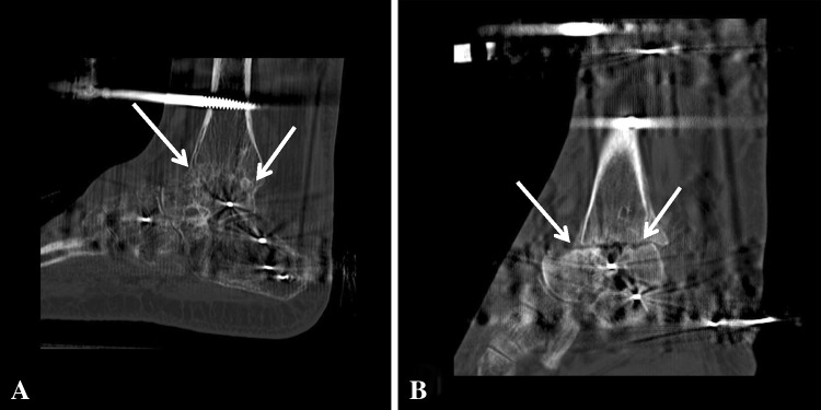
(A) The arrows highlight bridging bone across the tibiotalar fusion site as viewed on a CT scan taken 3 months after fusion. This image shows greater than 30% fusion healing, and the patient achieved successful union without further intervention. (B) No bridging bone across the tibiotalar fusion site (arrows) is seen at 3 months after fusion in a different patient.