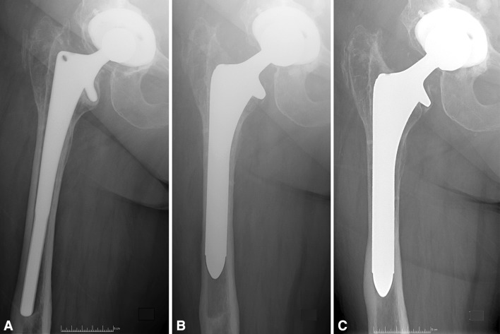
Fig. 2A–C.
(A) A preoperative AP radiograph demonstrates a loose cementless stem with a Paprosky Type IIIA femoral defect. (B) A 4-month postoperative AP radiograph shows the 6-inch fully porous-coated stem used at revision. (C) At 4-year followup, an AP radiograph shows that the stem remains well fixed with distal spot welds evidencing bone ingrowth.

