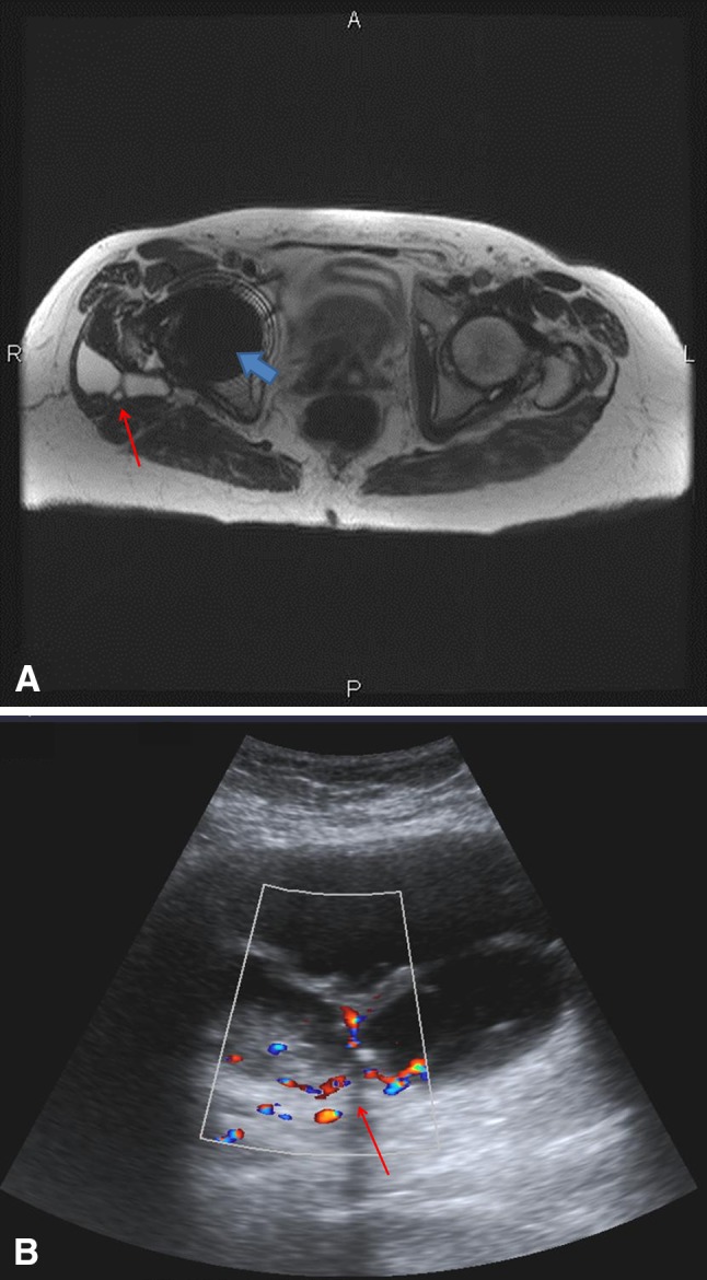
Fig. 1A–B.

MRI and ultrasound show a large pseudotumor in a 64-year-old woman. (A) A T2-weighted axial MR image using the SEMAC protocol shows a complex high T2 signal lesion (red arrow) posterolateral to the right femoral neck component of the MOM THA. The blue arrow demonstrates residual metallic artifact from the femoral head component of the THA. (B) A color Doppler ultrasound image in the axial plane shows a septated complex fluid collection (arrow) with internal echoes and vascularity within the septations, as shown by the color flow.
