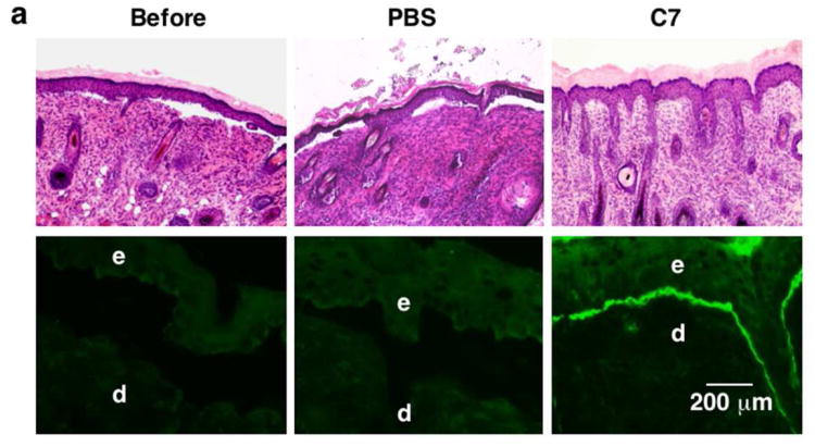
Figure 2. IV injected rC7 incorporates into the DEJ of RDEB mouse skin grafted onto athymic nude mice in vivo.
(a) Histological appearance (upper panels) and immunofluorescence staining (lower panels) of engrafted RDEB mouse skin using a monoclonal antibody specific for human C7. Left panels (Before) are skin biopsies from transplanted RDEB skin grafts taken two weeks after grafting and before treatment (n = 30 mice). Middle panels (PBS) are two-week post injection biopsies from RDEB skin grafts on athymic host nude mice that were IV injected with PBS (n = 4 mice). Right panels (C7) are two-week post injection biopsies from RDEB skin grafts on athymic host nude mice who were IV injected with 60 μg of rC7 (n = 9 mice). e, epidermis; d, dermis. (b) Immunogold labeling of engrafted murine RDEB skin injected with PBS (PBS) or 60 μg rC7 (C7) was performed with an antibody specific to human C7 (NP185, a gift of Dr. Lynn Sakai, Shriners Hospital for Children, Portland, Oregon) followed by an 1 nm gold secondary antibody. Identical IEM was performed on normal mouse skin (NMS). Note that IV injected rC7 incorporated into the RDEB skin grafts and formed AFs. Note restoration of numerous arching AFs depicted with arrows and labeled with gold particles decorating the DEJ of RDEB skin grafts received IV rC7. IEM on normal mouse skin shows numerous unlabelled AFs. HD = hemidesmosomes; D = dermis; E = epidermis; Scale bar: 100 nm.


