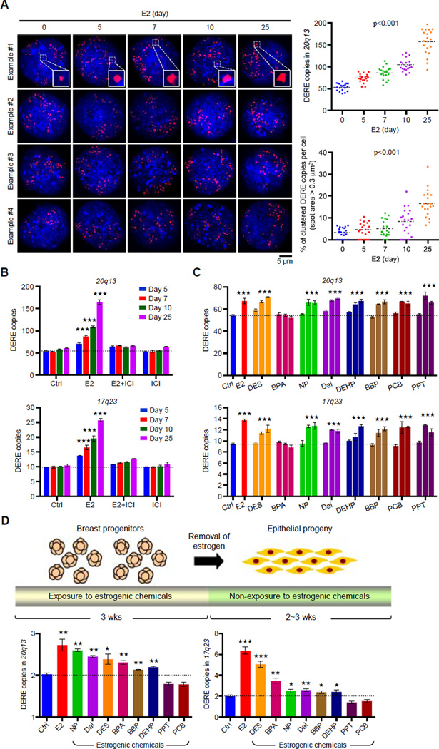
Figure 3. Prolonged Estrogen Exposure Leads to Amplification of DERE Copies.
(A) Interphase fluorescence in situ hybridization (FISH) analysis of amplified 20q13 DERE copies in E2-treated (70 nM) MCF-7 cells for different time periods (0, 5, 7, 10, and 25 days). Representative four images in each condition were shown. Inserted squares: clustered DEREs. Quantification of DERE copies per cell was performed by CellSens software and presented in the scatter plot (n=20). A spot with area size over 0.3 µm2 was counted as the clustered DERE region. p<0.001 (two way ANOVA test), compared to “0” group.
(B) Quantitative PCR analysis of two amplified DERE copies located in 20q13 (upper) and 17q23 (lower). MCF-7 cells were continuously exposed to E2 (70 nM) and/or ICI 182,780 (100 nM) for different time periods (5, 7, 10, and 25 days) in charcoal-stripped conditions (n=6 replicates in two biological batches of treatment). Mean ± SD. ***, p<0.001 (Student’s t test), compared to “Ctrl” cells.
(C) Dose-dependent gains of 20q13 and 17q23 DERE copy in MCF-7 cells exposed to different estrogenic chemicals. MCF-7 cells were cultured in charcoal-stripped conditions and exposed to ethanol (Ctrl), E2 (70 nM), or estrogenic chemicals with 5-fold different dose, including diethylstilbestrol (DES, 14-70-140 nM), bisphenol A (BPA, 0.5-4–20 nM), 4-nonylphenol (NP, 0.2-1–5 µM), daidzein (Dai, 2-10–50 µM), N-butyl-benzyl phthalate (BBP, 2-10-50 µM), di(2-ethylhexyl)-phthalate (DEHP, 2-10–50 µM), 4,4’-dichloro-biphnyl (PCB, 0.02-0.1–0.5 nM), and 1,3,5-tris(4-hydroxyphenyl)- 4-propyl-1H-pyrazole (PPT, 0.02-0.1–0.5 nM), respectively, for 5 days. These treatment doses were selected and modified based on our previous findings (Hsu et al., 2009 and 2010). Genomic DNA from treated cells was collected for quantitative PCR analysis of 17q23 and 20q13 DERE copies. Mean ± SD (n=6 replicates in two biological batches of treatment). ***, p<0.001 (Student’s t test), compared to “Ctrl” cells.
(D) Differential copy changes of 20q13 and 17q23 DEREs in normal epithelial cells preexposed to estrogenic chemicals. Experimental scheme of an in vitro exposure system is shown in the upper panel. Floating mammospheres containing breast progenitor cells were preexposed to dimethyl sulfoxide (DMSO as control, Ctrl), E2 (70 nM), or estrogenic chemicals, including DES (70 nM), BPA (4 nM), NP (1 µM), Dai (10µM), BBP (10µM), DEHP (10µM), PCB (0.1 nM), and PPT (0.1 nM), respectively, for 3 weeks. Differentiated epithelial cells were then subjected to quantitative PCR analysis (lower) of 17q23 and 20q13 DERE copies. Mean ± SD (n=6 replicates in two batches of treatment). ***, p<0.001 (Student’s t test), compared to “Ctrl” cells.

