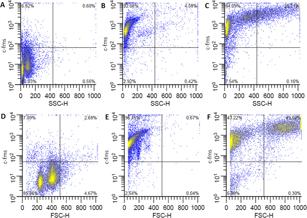
Figure 7. Flow cytometric analysis of c-fms on bone marrow derived macrophages.
Representative plots of c-fms fluorescence vs. side scatter (top row) or forward scatter (bottom row). Undifferentiated, freshly harvested bone marrow cells(A, D), or cells differentiated in medium containing 15 ng/ml M-CSF in FPAs (B, E) or Bioreactors (C,F). Culture vessels were incubated in horizontal orientations.

