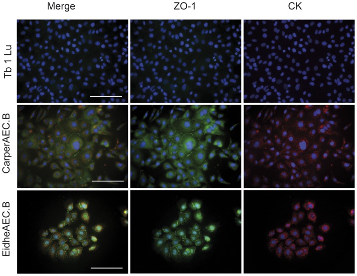
Figure 3. Immunofluorescence staining for markers of epithelial origin of Tb 1 Lu and airway epithelial cells from C. perspicillata (CarperAEC.B) and E. helvum (EidheAEC.B) prior to subcloning.
The markers used to confirm epithelial origin were cytokeratin (CK, red) and zonula occludens-1 (ZO-1, green); nuclei are counterstained with DAPI (blue). Expression of both markers is present in all cell lines generated by the described methods, indicating an epithelial origin. By contrast, the commercially available Tb 1 Lu does not show expression of the respective markers.

