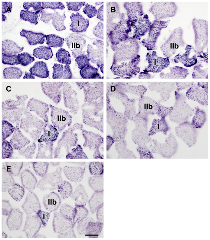
Figure 1. NADH-tetrazolium reductase stained sections from the ischemic experiment.
The figure shows representative pictures taken from the anterior tibialis muscle of untreated control animals (A), as well as after 4 (B), 6 (C), 8 (D) and 9 hours (E) of ischemia (induced by infrarenal aortic occlusion) without reperfusion. Typical members of Type I and Type IIb fibers are marked on each picture as I or IIb, respectively. Bar: 35 µm.

