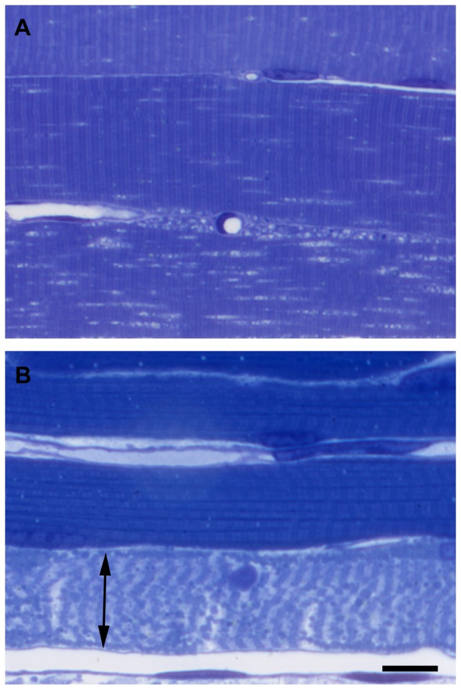
Figure 6. Morphological signs of ischemic-reperfusion damage of muscle fibers as demonstrated on semithin sections.

Marked morphological changes were visible in all fibers even after 8(A), while after 9 hours of ischemia followed by 2 hours of reperfusion (B) necrosis (double arrow) was already evident in the majority of the fibers. Toluidine blue staining. Bar: 20 µm.
