FIGURE 8:
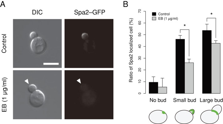
Effect of EB on Spa2–GFP localization. (A) YOC5002 (spa2 [Spa2–GFP]) cells were incubated at 25ºC in SD–U medium with EB (1 μg/ml) or DMSO (control solvent) until the early log phase. Cells were then harvested and observed without fixation. The arrowhead indicates the absence of Spa2–GFP localization in the bud. Bar, 5 μm. (B) Spa2–GFP localized cells were enumerated in three independent experiments. The mean of the triplicates is plotted. Error bars denote 1 standard deviation. Asterisk indicates significant difference (p < 0.05 by t test after Bonferroni correction).

