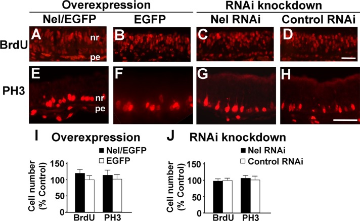
FIGURE 6:
Effects of Nel on proliferation of retinal progenitor cells. Expression constructs for Nel cDNA (A, E) or artificial miRNA (C, G) were transfected into the optic vesicle by in ovo electroporation at HH9–11 (E1.5), and effects on cell proliferation were examined by comparing with corresponding areas transfected with control vectors (EGFP in B, F, I; control RNAi in D, H, J). Areas in the central retina are shown. (A–D) Embryos were labeled with BrdU in ovo for 3 h at E6, and retinal sections were prepared and stained with anti-BrdU antibody. (E–H) Retinal sections of E4.5 chicks were stained for PH3. Scale bars, 50 μm. nr, neural retina; pe, pigment epithelium. (I, J) Quantifications of BrdU- and PH3-positive cells. The numbers of stained cells are shown as percentage control in mean ± SEM. No significant differences were detected between Nel overexpression, RNAi knockdown, and their controls. n = 6 embryos. ANOVA test.

