FIGURE 1:
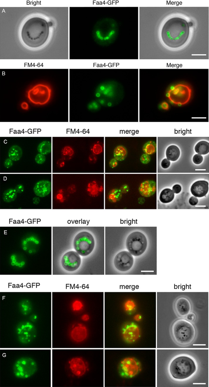
Lipid droplet–vacuole interaction and uptake in glucose- and oleate-grown yeast cells. LDs are labeled with endogenously expressed Faa4-GFP in cells grown on 0.5% glucose for 21 h (A) and 46 h (B). LDs are typically localized in strings adjacent to the vacuole (A) or randomly distributed in the cytosol. They are also frequently observed inside the vacuole, especially in the stationary phase of growth (absence of glucose; B). Cells expressing Faa4-GFP were pregrown on glucose and subsequently shifted to oleate-containing media. After 6 (C) and 12 (D) h of incubation, LDs are massively induced in the cytosol and are also present inside the vacuoles. In stationary phase (28 h of incubation) distinct LDs are no longer detectable in the vacuole (E). After shift of these cells to fresh oleic acid–containing medium lacking a nitrogen source, LDs are rapidly incorporated into the vacuole: after 1 h (F) and 5 h (G). Vacuolar membranes are stained with FM4-64. Scale bar, 5 μm.

