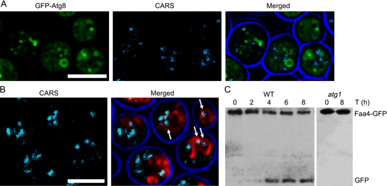
FIGURE 3:
Lipid droplets are degraded in the yeast vacuole upon induction of autophagy. (A) ypt7 cells expressing GFP-Atg8 show the accumulation of autophagosomes that lack LDs. (B) Detection of LDs inside the vacuole of wild-type cells with CARS imaging; vacuolar membranes are labeled with FM4-64. Cells were shifted to nitrogen starvation medium for 8 h in the presence of PMSF before microscopy to induce autophagy. Scale bar, 5 µm. (C) Western blot of cell extracts of wild-type cells expressing the LD marker Faa4-GFP, using an anti-GFP antibody. Late exponential cells grown in rich medium were shifted for 8 h to medium lacking a nitrogen source. The appearance of one or two bands at ∼27 kDa is indicative of vacuolar proteolytic processing of the Faa4-GFP fusion protein. This band is absent in atg1 cells.

