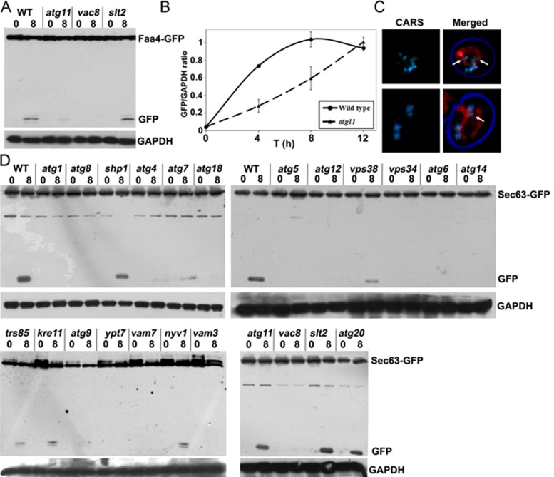
FIGURE 6:
Lipid droplet autophagy requires selective adapters and differs from ER-phagy. (A) Protein extracts of various mutant cells expressing Faa4-GFP were grown to the late logarithmic growth phase in rich medium and shifted to synthetic minimal medium lacking nitrogen for the indicated time intervals. This analysis shows the requirement for Vac8 and a partial requirement for Atg11 for Faa4-GFP cleavage. Blots were decorated with anti-GFP and anti-GAPDH antibodies. (B) Quantification of cleaved Faa4-GFP at different time points after the shift to starvation medium in wild-type and atg11 mutant cells expressing Faa4-GFP relative to the GAPDH loading control. (C) CARS images of atg11-mutant cells shifted to nitrogen starvation medium for 8 h in the presence of PMSF. LDs are internalized into vacuoles of atg11 cells that are labeled with FM4-64. (D) Protein extracts from various mutant cells expressing the ER marker Sec63-GFP analyzed by Western blotting. Cells were grown to the late logarithmic growth phase in rich medium and shifted to synthetic minimal medium lacking nitrogen for indicated times. Blots were decorated with anti-GFP and anti-GAPDH antibodies. This analysis shows that LD autophagy is distinct from ER-phagy. See the text for details.

