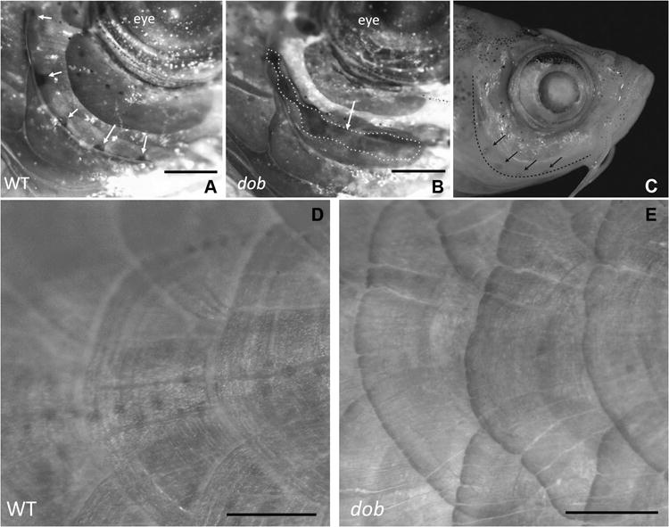
Figure 4.

The effect of the loss of fgf20a expression on zebrafish mineralized tissue anatomy. A. Lateral view of a ABWT specimen with normal lateral line formation in the preopercle bone. Lateral line pores are indicated by arrows. B. Lateral line canals are incompletely fused in dob fish, resulting in gaping clefts in the canals (dotted line/arrow). For A and B dorsal is up and anterior is to the right. C. ABWT zebrafish head showing the edge of the preopercle bone (dashed line) and lateral line pores in this bone (arrows). D. Normal body scales in a ABWT fish. E. Distorted body scales with irregular posterior margins in a dob fish. The scales depicted in D and E are located along the posterior flank of the fish. Dorsal is up and anterior is to the right. Scale bars equal 500μm.
