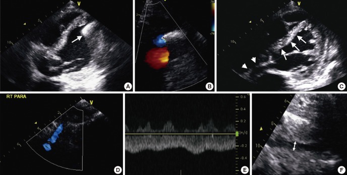
Fig. 2.
Checkpoint in echocardiographic assessment after LVAD implantation. (A, B) The axis and laminar flow of inflow conduit in modified apical-4-chamber view, (C) The position of interventricular septum (IVS) and interatrial septum (IAS), (D, E) The flow of inflow cannula in right parasternal view, and (F) IVC diameter in subcostal view.

