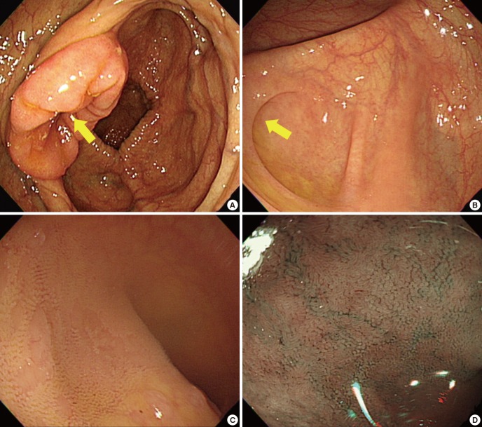
Fig. 3.
Colonoscopic landmarks in photographic documentation of complete cecal and terminal intubation. (A) Ileocecal valve (yellow arrow). (B) Appendiceal orifice (yellow arrow). (C) Terminal ileum. Villi were seen in the terminal ileum (water-filling method). (D) Terminal ileum. Villi were seen in the terminal ileum (narrow-band imaging method).

