Figure 3.
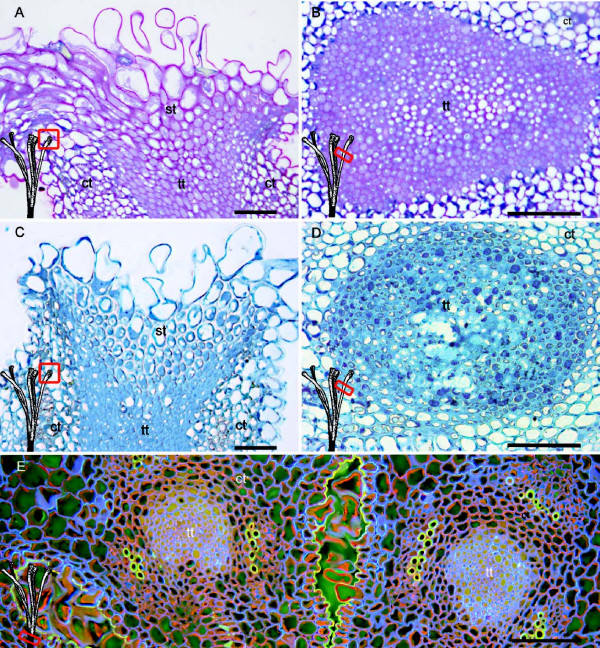
Stigma and stylar transmitting tissue of Malus x domestica at anthesis. (A) Stigma and stigmatoid tissue with intercellular spaces filled with insoluble polysaccharides after staining with Periodic acid Shiffs-PAS reagent counterstained with toluidine blue. (B) The extracellular matrix of the transmitting tissue also stained for polysaccharides. (C) Absence of general protein staining with Naphtol Blue Black in the intercelullar space of the stigmatoid tissue, contrasting with (D) strong reaction both within cells and in the intercellular space of the style transmitting tissue with the same staining. (E) Reduced area of the transmitting tissues at the stylar base in the multicarpelar apple pistil stained with acridine orange counterstained with aniline blue. Light micrographs of 2 μm longitudinal stigmatic sections (A,C), and transversal style sections in the upper style (B,D - red squares), and at the stylar base (E-red square). Cortical tissue (ct), stigmatoid tissue (st), transmitting tissue (tt). Scale bars: 50 μm.

