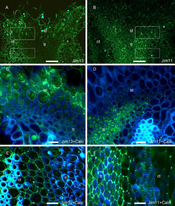
Figure 5.
Glycoproteins in the stigmatoid and stylar transmitting tissue of Malus x domestica, at anthesis. (A) Arabinogalactan proteins labelled with JIM13 mAb were present in the cortical and stigmatoid tissues, but were absent in the stylar transmitting tissue. (B) In contrast, extensin epitopes were conspicous in the transmitting tissue of the style. (C) While the stigmatoid tissue intercellular space positively reacted for the presence of arabinogalactan proteins epitopes recongized by JIM13 mAb, (D) intercellular spaces of the contiguous transmitting tissue was marked by the presence of extensins. (E) The upper style further lacked JIM13 arabinogalactan protein epitopes in the transmitting tissue. (F) In contrast the intercellular space in the style transmitting tissue contained extensins. Longitudinal 4μm sections of the stigma-style transition (A-D), and the style (E-F), tagged either for arabinogalactan proteins with JIM13 mAb (A,C,E), or extensins with JIM11 mAb (B,D,F). Merged images of FITC labelling (green) with calcofluor white (blue) (C-F). White squares note the location of the magnified C,E and D,F pictures in each column respectively. ct, cortical tissue; st, stigmatoid tissue; tt, stylar transmitting tissue. A-B scale bars: 50 μm; C-F scale bars: 10 μm.

