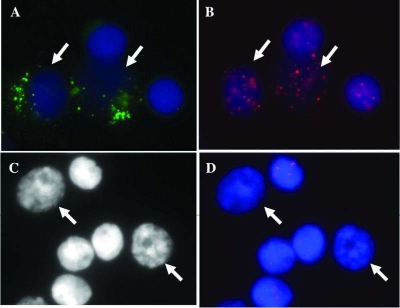
Figure 1.
Telomere signals in CD138+ plasma cells (A–D). (A) Myeloma cells fluoresce green, whereas normal cells remained unstained (see arrows). (B) The telomeres, hybridized with Cy3-labeled PNA probes, appear as red signals. The nuclei are counterstained with DAPI (blue). (C) Identification of 3D fixed nuclei in myeloma cells and normal lymphocytes based on size and intensity of the counterstain DAPI (see arrows). (D) Cy3-labeled PNA telomeres in 3D fixed lymphocytes.

