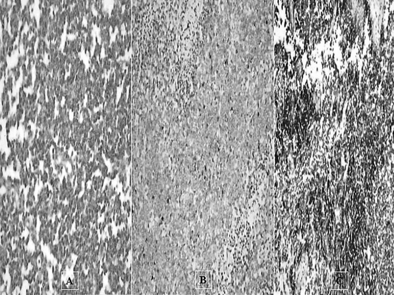
Figure 3.
Biopsy sample pathology (A) High power magnification of the small cell carcinoma portion (H&E stain ×200). Note that the tumor cells are often oat shapedomatin with inconspicuous nucleoli. (B) The tumor cells are negative for LCA immunostaining, (C) positive for Synaptophysin immunostaining.

