Fig. 5.
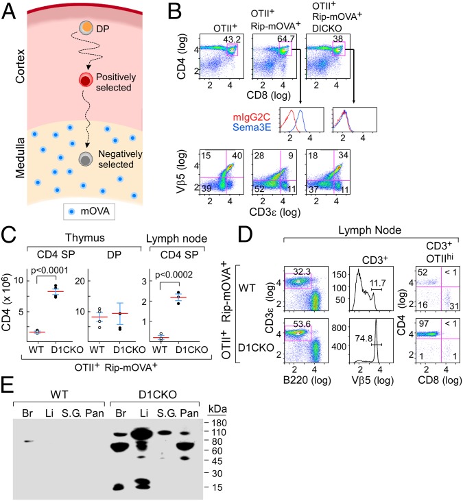
Loss of thymocyte plexinD1 leads to defective negative selection. (A) Schematic of OTII+RIP-mOVA+ thymocyte selection. Ovalbumin expressed from the Rip promoter is degraded in the thymic medulla and presented on mTEC (blue) to positively selected thymocytes migrating from the cortex. For negative selection, OTII-TCR+ thymocytes must move to the medulla. (B) Survival of CD4+ SP OTII+RIP-mOVA+ D1CKO thymocytes in comparison with plexinD1-expressing OTII+RIP-mOVA+ thymocytes. (Top) CD4 vs. CD8 FACS dot plots on total thymocytes from indicated mouse strains. (Middle) Expression of plexinD1 enumerated by sema3E-Fc binding on gated DP thymocytes. (Bottom) Enumeration of mature Vβ5/CD3hi thymocytes. (C) Survival of thymic and lymph node CD4 SP cells (absolute number per organ). Mean ± SD is shown. (D) Survival of OTIIhi lymph node T cells, gating on CD3ε then Vβ5 to observe CD4 subset. (E) Serum autoantibody reactivity with different tissues as detected by Western blotting (Br, brain; Li, liver; S.G., salivary gland; Pan, pancreas). Results of individual panels are representative of two to three independent experiments for each group of mice examined (where total n > 8).

