Fig. 4.
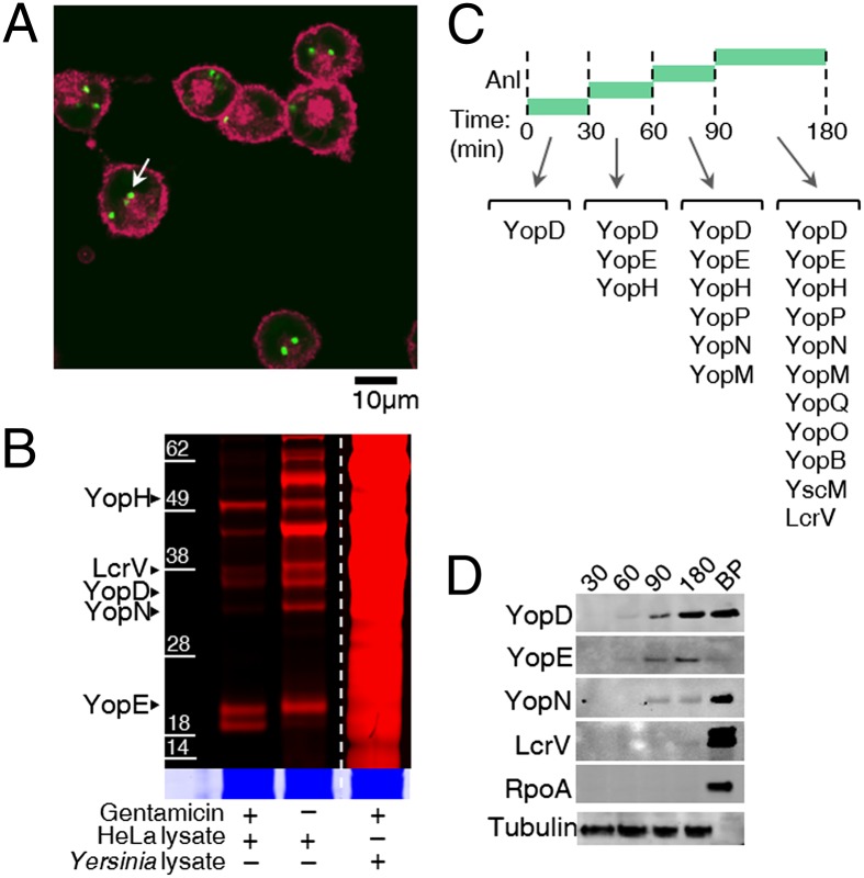
Labeling of proteins injected into HeLa cells by internalized Y. enterocolitica and identification of the order of Yop injection into HeLa cells. (A) Confocal fluorescence microscopy showed Anl incorporation into the proteome of internalized Y. enterocolitica. HeLa cell membranes were labeled with Alexa Fluor 633-WGA conjugate (red), and Anl residues were labeled with alkyne-Alexa Fluor 488 (green). The arrow indicates labeled Y. enterocolitica inside HeLa cells. (B) Infected HeLa cells were selectively lysed with digitonin and treated with alkyne-TAMRA to detect the proteins injected by internalized Y. enterocolitica. (Inset) Colloidal blue staining of the same gel. In the presence of gentamicin only, internalized Yersinia can inject proteins into HeLa cells. (C) The order of injection of Yops was determined by pulsed-Anl labeling and shotgun MS. Anl was added only during the indicated times for each infection, and HeLa cells were lysed with digitonin at the end of each time interval. (D) Western blot analysis detected Yops in pulsed-Anl labeling experiments. RpoA served as a control for bacterial lysis; antibody for α-tubulin was used as a loading control for HeLa lysates. BP, bacterial pellet.

