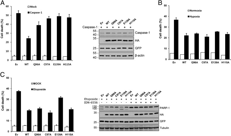
Fig. 5.
Evaluation of APIP/MtnB enzymatic mutants for the inhibition of cell death. (A) Caspase-1–induced cell death. (Left) HeLa cells were transiently cotransfected with pEGFP and the indicated constructs for 18 h. After staining with 0.5 µg/mL ethidium homodimer (EtHD), cell death rates were measured by counting the number of both GFP- and EtHD-positive cells among total GFP-positive cells after staining with EtHD. Ev, empty vector. Values indicate mean ± SD (n = 3). (Right) Whole-cell extracts were prepared and subjected to Western blotting using the indicated antibodies. (B) Hypoxia-induced cell death. HeLa cells were transiently cotransfected with pEGFP and the indicated constructs for 24 h and then exposed to a hypoxic condition of 1% O2 for 48 h. Cell death rates were measured by trypan blue exclusion assay; bars represent mean ± SD (n = 3). (C) Etoposide-induced cell death. (Left) HeLa cells were transiently cotransfected with pEGFP and the indicated constructs for 24 h and then treated with 40 μM etoposide for 36 h. After staining with 0.5 μg/mL EtHD, cell death rates were measured by counting the number of both GFP- and EtHD-positive cells among total GFP-positive cells. Bars indicate mean ± SD (n = 3). (Right) Cells were treated the same as described above in the presence or absence of 10 μM IDN-6556. Western blotting was performed with the indicated antibodies. PARP-1, poly(ADP ribose) polymerase-1.

