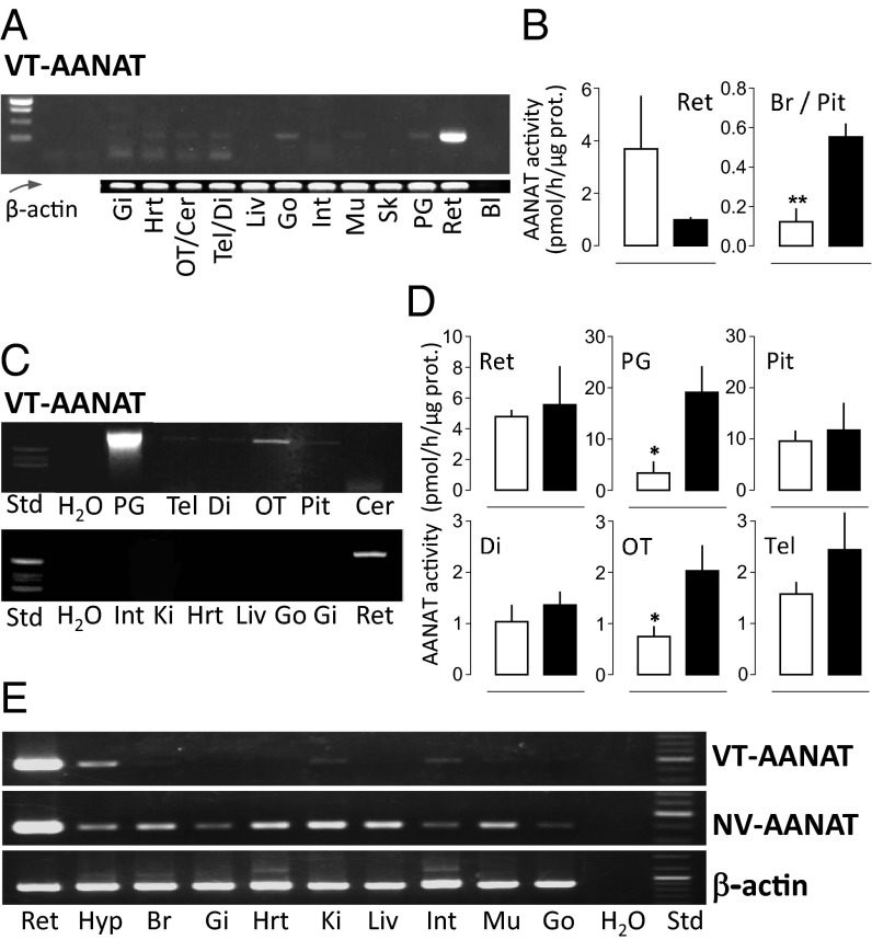
Fig. 2.
Tissue distribution of VT-AANAT in lamprey, catshark, and elephant shark. (A, C, and E) Presence of VT-AANAT mRNA in (A) lamprey, (C) catshark, and (E) elephant shark tissues as indicated by RT-PCR. (B and D) VT-AANAT activity in (B) lamprey and (D) catshark neural organs and tissues. Sampling was performed at midday (white bars) and midnight (black bars). Mean ± SEM (n = 5). Student t test: *P < 0.05; **P < 0.01. Additional details in SI Appendix. Bl, blank; Cer, cerebellum; Di, diencephalon; Gi, gills; Go, gonads; Hrt, heart; Hyp, hypothalmus; Int, intestine; Ki, kidney; Liv, liver; Mu, muscle; OT, optic tectum; Pit, pituitary; PG, pineal gland; Ret, retina; Sk, skin; Tel, telencephalon.

